Friday, January 28, 2011

Fistula

Definition of Fistula:
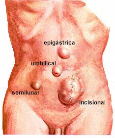
Fistula is defined as an abnormal tract lined by epithelium,endothelium,granulation tissue or fibrous tissue connecting two epithelial surfaces either in between the exterior and an internal viscus(External fistula) or in between two internal viscera(Internal fistula).

Classification:


A.Congenital:
  • Bronchial fistula(neck,lower 1/3rd)
  • Tracheo-oesophageal fistula
B.Acquired:
  1. Fistula-in-ano
  2. Arteriovenous fistula-Hemodialysis,Trauma
  3. Neoplastic
  • Rectovaginal fistula
  • Gastrocolic fistula

Anorectal fistulas


    Clinical features of Sinus/Fistula: 
    1.Persistent or intermittent discharge from an abnormal opening.
    2.intermittent swelling and pain of the local site followed by discharge and relief of pain.
    3.Symptoms of underlying pathology,e.g.,
         a)TB
    • Low grade fever
    • Cough
    • Haemoptysis
    • Chest pain
    • Dyspnoea
         b)Congenital sinus/fistula:
    From birth,e.g.,Preauricular sinus
         c)Osteomyelitis(OM)
    • High fever
    • Pain
    • Swelling of the bone concerned
         d)Perianal sinus/fistula:
    History of ischiorectal abscess

    Causes of persistence of a sinus:
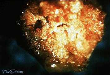
    1.Presence of FB or necrotic tissue
    • Suture material
    • Sequestrum(Necrotic tissue and foreign body)
    • Bullet
    • Pellet
    • Worms
    2.Non-dependent drainage or inadequate drainage
    3.Abscess of rest for
    • Fistula in ano(Proximal colostomy is done)
    4.Distal obstruction
    5.When the tract is lined by epithelium or endothelium
    6.When the tract contains excessive fibrous tissue(prevents collapsing) around the tract(e.g.,Chronic empyema chest)
    7.Naturre of infection
    • TB
    • Actinomycosis
    8.Underlying malignancy
    9.Persistent discharge(VVF-urine)
    10.Chronic illness or systemic illness
    • DM
    11.Immunosuppression
    12.Nutritional deficiency
    13.Drugs
    • Steroid
    14.Irradication:
    • RVF in treating carcinoma of cervix
    15.Ischaemia

    Principle of Treatment of Sinus/Fistula:
    1.Excision followed by biopsy
    2.Treatment of the underlying cause(e.g., TB,DM,Carcinoma,Malnutrition)

    Peptic Ulcer

    Definition of Peptic Ulcer: Peptic ulcers are chronic, most often solitary lesion occuring in any part of the GIT exposed to the aggressive action of acid peptic juices.

    Or
    It is a chronic ulcerative lesion of the GIT at sites exposed to aggressive action of acid peptic juices.


    Peptic ulcer


    Types of Peptic Ulcer:
    • Duodenal Ulcer
    • Gastric Ulcer
    Sites of ulcer in GI tract:
    1. Commonest site:First part of the duodenum on the anterior wall(about 70%)
    2. Within the antral part of stomach towards the lesser curvatures close to the junction of acid secreting and non-acid secreting part.(About 28%)
    3. Lower end of the esophagus (Barrett's esophagus,a site of columnar metaplasia)
    4. Stomach ulcer,within the margins of a gastrojejunostomy.
    5. In the duodenum,stomach or jejunum of patients with Zollinger Ellison Syndrome:Non -B cell of pancrease producing gastrin.This gastrin causes hyper secretion of gastric acid.This tumor may found in any part of GIT.
    6. Mickle's diverticulum that contains ectopic gastric mucosa.
    Risk factors/Causes of peptic ulcer:
    Imbalance between gastric mucosal barrier (defense mechanism) and acid pepsin mixture(damaging forces):
    1. Hyper acidity
    2. Mucosal barrier defect(in gastric ulcer)
    3. Predisposing condition:-
              a)Genetic influence(duodenal ulcer)

    • Family
    • 'O' negative blood group.
              b)Environmental
         4.H.pylori infection (Chronic gastritis)-About 85-100% of patients with duodenal ulcer & 65% with gastric ulcer.

    Treatment of peptic ulcer:

    A.Relieve pain by raising gastric pH(alkalization)
    • Neutralization of gastric acid by antacids.
    • Inhibition of gastric secretion by-H2 blocker,Anticholinergic drugs,PGE2
    • Surgical removal -Gastrectomy
    B.To prevent complication
    C.To promote healing
    • Bed rest & sedation-Diazepam]
    • Use of ulcer healer drugs-sucralfate
    D.To prevent recurrence-
    • Regular diet
    • Avoid ulcerogenic drugs-Aspirin,Steroids
    • Avoid smoking,alcohol,tea,coffee.

    Wednesday, January 26, 2011

    Hernia

    Definition:
    It is an abnormal protrusion of a part or whole of a viscus through an opening in the wall of the cavity in which it is contained.

    Types of hernia:
    A.External:
    1. Inginal
    2. Umbilical
    3. Femoral(female)
    4. Incisional
    B.Internal:
    1. Hiatus hernia
    2. Herniation of brain through the foramen of morgagni
    3. Herniation of abdominal organ through the diaphram.
    Commonest:Indirect inguinal hernia(commonest of all)
    Commoner hernias:
    1. Inguinal
    2. Femoral
    3. Incisional
    4. Umbilical
    Clinically 4 types:
    1. Reducible
    2. Irreducible
    3. Obstructed
    4. Strangulated
    Parts of hernia: Herniaa consists of three parts
    1.Sac:
    • Mouth
    • Neck
    • Body
    • Fundus
    2.Contents:
    • Intestine-enterocele
    • Omentum-Omentocele
    Hernia pictures:


    Hernia pictures of male genitalia:


      Symptoms of hernia:
      1.A lump
      2.Discomfort of pain-due to stretching of neck.
      severe pain-obstruction/strangulation
      3.Vomiting

      Treatment of hernia:
      • Indirect inguinal hernia(Uncomplicated)
      Adolescent,Young adult-Herniotomy
      They have very good inguinal muscle.
      Elderly-Herniotomy and herniorrhaphy.
      • Complicated(strangulated)
      Emergency operation done after resuscitation.

      Buerger's Disease

      Definition:
      It is a progressive,peripheral,vascular occlusive disease due to involvement of all the three layers of small to medium sized arteries in an non specific inflammatory process.

      Features:

      1.Occlusive disease of medium and small sized arteries:
      • Digital
      • Planter
      • Dorsalis pedis
      • Anterior tibial
      • Radial
      2.Thrombophlebitis of superficial and deep veins.
      3.Raynaud's phenomenon:In male patient in younger age group.

      Symptoms:
      1.Pain in the limb
      a)Intermittent claudication
      b)Rest pain
      Tingling and numbness/Paresthesia
      • Loss of sensation
      • Coldness of the part
      • Color changes
      • Ulceration or combination of the affected part
       Buerger's Disease pictures:


        Sign:

        A.General signs:
        1.patient is anxious
        2.Patient prefers to sit by the side of the bed hanging the limb,unwilling to lie flat

        B.Local signs:
        • Inspection:
        1. Color changes with line of demarcation
        2. Loss of hair
        3. Skin-dry,shrivelled,mummified,blackened,greasy to touch
        4. Ulcer (if any) examine accordingly
        5. Nails:falling or trophic changes.
        6. Muscle wasting.
        • Palpation:
        1. Temp:cold
        2. Pulse:lost
        3. Sensation:decreased
        4. Motor power:decreased
        5. Reflex function:decreased
        6. Buerger's test
        7. Buerger's angle.
        • Auscultation:
        1. Bruit
        C.Systemic examination: CVS

        Treatment:
        A.General:
        1. Stop smoking:prevents progress of disease
        2. Nutritious diet and vitamin supplements
        3. Care of the foot/feet:wearing soft shoes and socks,keeping the feet dry,Careful paring of toe nails
        4. Regular exercise within the limit of claudication pain(distance)
        5. Vasodilator drugs:Nicotinic acid,Isosorbide dinitrate
        6. Analgesics:Diclofenac
        B.Specific Rx:Depends on the condition/situation

        Monday, January 24, 2011

        Sexually Transmitted Disease(STDs)

        What is STD: Sexually transmitted disease (STDs) are agroup of communicable diseases that are transmitted predominantly by sexual contact and caused by a wide range of bacterial,viral,protozoal & fungal agents & ectoparasite.

        Classification of STDs:

        1.Disease manifested by discharge-
        • Gonorrhea(N.gonorrhoae)
        • Non gonococcal urethritis
        2.Disease manifested by ulcer-
        • Syphilis(T.pallidum)
        • Herpes and cervical cancer(HHV-2 & HHV-1)
        • Chancroid (H.ducreyi)
        • Granuloma inguinale
        3.Disease manifested by lumps-
        • Lymphogranuloma venereum(Chlamydia trachomatis)
        • Molluscum contagiosum(one of the pox virus)
        • Genital warts(Papilloma virus)
        4.Vaginitis-

        • Trichomoniasis(T.vaginalis)
        • Vaginal thrush(Candida albicans)
        • Bacterial vaginitis(Gardnerella vaginalis)
        5.Infestation with parasite-
        • pediculosis pubis
        • Genital scabies(Sarcoptes scabei hominis)
        6.AIDS- HIV-I,HIV-II


        7.Hepatitis virus- HBV,HCV,CMV

        8.PID,Nonspecific urethritis,Proctitis,Cervicitis- Bacterial infection (Chlamydia,Mycoplasma, hominis,urea plasma)

        9.Additional infection in homosexual man-
        • Bacterial: Shigella,Salmonella,Campylobacter jejuni
        • Viral: HIV-I,HIV-II,HBV,HDV,HCV,HAV,CMV
        • Protozoal: Giardia,E.histolytica,Cryptosporidium and Microsporidium.
        • Helminthic: Enterobius vermicularis,Trichuris trichuria,Strongyloides stercoralis.
        Investigation of STD:
        1.Material collection
        a.urethral specimen or swab in urethritis
        b.Vaginal swab in vaginitis
        c.Cervical swab in women with or without puerperal sepsis
        d.Fluid and pus from genital ulcer

        2.Laboratory examinations:
        Examinations are to be planned on clinical diagnosis-

        A.First day-
        • Microscopic examination of the specimen,Gram stain of the smear
        • Culture of the specimen
        B.Second day & onwards-

        Serology:
        1.Test for detection of HIV:
        • ELISA-screening test
        • Western blot-confirmatory test
        2.Test for detection of HBsAg-(ELISA) ,Haemagglutination test.

        3.Test for syphilis:
        • VDRL test
        • Complement fixation test
        • Kahn test
        4.Antitreponemal  antibody tests:
        • TPHA
        • FIA-ABS test
        5.Other tests:
        • Gonococcal CFT
        • Herpes CFT
        • Chlamydia CFT

        Friday, January 21, 2011

        Obsessive-compulsive disorder

        Obsession:Obsession is a recurrent and intrusive thought,emotion,impulse and idea that comes into patients mind against his or her will.
        It may be unrealistic.It will produce anxiety.

        Compulsion:It is an act or ritual which patient does not get relief from anxiety e.g washing of hand when patient thought it is dirty.
        Compulsion always follows obsession.There may be obsession only.

        Epidemiology:
        2-3% life time prevalence
        1% seriously disabling(hampering normal life)
        Peak age:25-30yrs.
        Female:Male=2:1
        In clinical practice F:M=1:1

        Causes:
        1.Biological factors:
        5-HT hormone is responsible for obsession.Decrease 5-HT level produce obsession.
        2.Psychological fctor:
              a)Psychoanalytic:Fraudian theory
              b)Behavioural:learning theory
        3.Genetic factors:genetic transmission-not strongly suggested,but having some weak relation.

        Clinical features:
        Some obsession is compatible with life process.Some ritual follows by some people. They do not come for the patient of moderate and severe feature usually come for treatment>

        1.Compulsion:
        a)Cleaning-most compulsion seen
        b)Counting:pt 2,4,6,8 count as like during passing chowkat,to see lamppost and count birds
        c)Checking:After locking the key,is it locked
        d)Dressing:Wear definite cloth after collecting all cloths

        2.Obsession:
        a)Thought-
        • Religious:God does not exist in those who prayer
        • Sexual
        3.Impulse:To destroy the patient or others e.g.to kill child by pressing neck,desire to jump from balcony
        4.Image:That are dirty e.g vomitus,stool-memorise during eating and vomiting
        5.Feeling:Dirty feeling about beloved person and emotion.

        If obsession persists for longtime,the patient develops depressive illness and may commit suicide.
        Sometimes the patients are so occupied with obsession that he neglect personal hygiene and develops-
        • Wt loss,cachexia
        • Insomnia
        • Low libido
        • Loss of appetite
        Anorexia nervosa:
        The patient does not eat or become so busy that did not get any time for eating.

        Management of Obsessive-compulsive disorder:

        1.Pharmacotherapy-
        TCA-
        • Chlomipramine:50-200mg/day
        • Fluoxetine:40-80mg/day to be taken in the morning
        Antipsychotic drugs may be added such as
        Trifluperazine:5-15mg/day

        2.Psychotherapy:
        Behavioural therapy:
        • Systemic desensitisation:done for response prevention
        • Thought stopping:disturbing thought think about that and stop that.
        Response is >95% if both methods are used.Alone therapy<80%
        If fails-Psychosurgery
        Sometimes before psychosurgery,electroconvulsive therapy.
        If treated early -good prognosis
        Discontinuation of therapy may lead to relapse of disease.
        But when untreated for 5 years ,it may run for life long.
        Good response if treated within 1-2 years. Patient may have co-morbid disorder with obsessive-compulsive disorder e.g.schizophrenia

        Tuesday, January 18, 2011

        HIV(Human Immunodeficiency Virus

        Properties of HIV virus:
        • HIV is a RNA virus belongs to the Retroviridae;was first described in 1981 and isolated in 1983.
        • HIV belongs to the lentivirus subgroup of retrovirus
        • Has double copy of the genome(diploid)
        • Possess reverse transcriptase enzyme
        • Has envelope
        • Surface antigen-glycoprotein120,glycoprotein 41
        • Incubation period-6 months to 6 years or more(Causes slow virus infection)
        • Both HIV-1 and HIV-2 causes AIDS,but HIV-1 is found worldwide,whereas HIV-2 is found primarily in west Africa.
        • HIV infects and kill helper T cell, resulting in the loss of cell mediated immunity, and causes opportunistic infection.Other cells e.g.macrophages and monocytes have CD4 proteins on their surface can be infected also.
        HIV model: virus structure


          Mode of Transmission of HIV:

          • Sexual contact with infected individual-vaginal sex, anal sex, deep kiss.
          • Transmission in blood:Receiving infected blood, blood products(Factor VIII concentrates.)
          • Sharing needles as in drug abusers or accidental inoculation.
          • Peripheral transmission:Mother to baby,either transplacentally(vertical) or perinatally. Infection may be acquired from breast milk.


            Pathogenesis of AIDS: After entry into blood stream,HIV attaches to CD4-(Helper T) cell & lymphocytes; grows slowly in CD4+ and monocytic cell. The deplition of CD4+ cells and macrophages result in impaired vell mediated immunity and humoral immune responses. The normal ratio of CD4+ to CD8+ is 2:1; which is reversed as 1:2. This leads to recurrent life threatening opportunistic infections.

            Clinical Features of AIDS:
            a.Early(acute) clinical features(begins after 2-4 weeks of infection):
            • Fever
            • Lethargy
            • Sore throat
            • Diarrhoea
            • Generalized lymphadenopathy
            • Maculopapular rash on the trunk,arms,and legs (but sparing the palm and sole)
            • Leucopenia occurs,but the number of CD4 cells is usually normal,high level of viremia occurs
            Antibodies to HIV typically appear 10-14 days after infection.
            b.Intermediate stage:
            AIDS related complex(ARC)-
            • Persistent fever
            • Fatigue 
            • Weight loss
            • Generalized lymphadenopathy
            c.Late stage of HIV infection is AIDS: Patient develops opportunistic infection by both pathogenic & non-pathogenic organisms.

            Laboratory diagnosis of AIDS: 
            AIDS is practically diagnosed by serological test for antibody against the virus.Electron microscopy is done in reference lab.Cell culture is done for research purpose.NA based techniques are used in special cases.

            Treatment of HIV infection(AIDS):
            A.Current treatment of choice:
            Regimen consisting of two-nucleoside inhibitor(Zidovudine & Lamivudine)-this combination is known as HAART [highly active antiretroviral therapy]
            B.In addition to nucleoside inhibitor there are non-nucleoside reverse transcriptase inhibitors that are effective against HIV-Nevirapine(viramune),delavirdine(rescriptor) & efavirenz(Sustiva)
            C.Protease inhibitor: Saquinavir(Invirase), ritonavir(Norvir),nelfinavir(viracept) & indinavir(Crixivan)

            GOZmosis:On Cure for HIV


            Prevention of AIDS:
            • Avoidance of promiscuity & anal intercourse
            • Use of condom
            • Screening of blood & blood products
            • Use of disposable syringe and needles
            • Interruption of perinatal transmission by screening women of child bearing age.
            • Counseling of person found to be antibody positive
            • Education about the mode of transmission & how to avoid infecting others & how to protect themselves.

            Wednesday, January 12, 2011

            Lymphogranuloma Venerum

            It is a STD caused by Chlamydia tracomatis(L1,2,3)

            Clinical features:
            Primary lesions are 2 to 3mm vesicles or erosions over genitalia
            Secondary-inguinal lymphadenopathy occurs after 2 weeks become swollen and tender
            Bubo-ulcerated
            Groove sign-due to swollen inguinal and femoral lymphnode(Inguinal ligament become tight-due to involvement of femoral and inguinal lymphnode)

            Lymphogranuloma Venerum pictures:


            Diagnosis:
            1. Giemsa stain-lymphnode aspirate isolation of LGV agent
            2. LGV-CFT
            3. Microhemoagglutination test-confirm species and strain (L1,2,3)
            Treatment:
            Doxycycline 100mg for 21 days or Eryrhromycin
            500mg q.d.s for 21 days

            Tuesday, January 11, 2011

            Carbuncle

            Definition of Carbuncle:
            Carbuncle is defined as the infective gangrene of the skin and subcutaneous tissue composed of boil with multiple formed or incipient discharging sinuses usually due to staphylococcus.(seen in nose following fever)

            Common sites:
            1.Nape of neck
            • Less vascular area
            • lax and extensive tissue planes
            2.Back  of the shoulder
            3.But can occur anywhere in the body

            Susceptible groups:
            1. Diabetic patients
            2. Usually male
            3. Age:more than 20 yrs
            4. Chronic illness or immunodeficiency.
            Complications:
            1. Septicaemia
            2. Epidural abscess
            3. Meningitis
            2,3 more common in nape of the neck.

            Management:
            1. incision and drainage of pus and excision of all dead tissue.
            2. Control of diabetes mellitus
            3. Antibiotics(cloxacillin or flucloxacillin)
            4. Regular local wound care(Dressing -in hospital,usually at 2 or 3 days interval)
            5. Improvement of the general condition of the patient.

            Monday, January 10, 2011

            Mumps

            Common in younger children(below 15yr)
            Young adult may also be affected at the dormitory,barracks etc.
            Mumps of mother during last trimester of pregnancy,there may be chance of mump infection in infant after birth,although he gets immunity.

            Mode of transmission:
            • Direct contacts
            • Droplets
            • Fomites
            Causative agent:Paramyxo virus
            May be endemic
            Immunity is life long but second attack may be possible(0.5%)

            Incubation period:14-24days.

            Clinical features:
            1. Fever
            2. Bodyache,malaise
            3. Swelling over parotid gland-first unilateral and involve other.Swelling completed within 2-3 days and subsides within 7 days.
            4. The swelling is painful.The swelling distorted the angle of mouth.It is associated with regional lymphadenitis.
            Diagnosis:
            1. Clinical
            2. No definite lab aid with limited facilities.The consequence of mumps infection are detroitus
            3. TC,DC:lymphocytosis,leukopenia
            4. Viral culture from blood and saliva
            5. Serological test
            Complications of mumps:
            1. Meningoencephalitis,symptoms of meningitis,altered consciousness,disorientation
            2. Orchitis and epididymitis
            3. Oophoritis
            4. Pancreatitis:serum amylase level increased
            5. Myocarditis
            6. Gastritis
            7. Thyroiditis
            8. Nephritis
            9. Unilateral deafness
            10. Occular manifestation-optic neuritis
            11. Dacryoadenitis
            12. Thrombocytopenia
            13. Arthritis
            14. Mumps embryopathy:developmental malformation of child if mother suffer from mumps infection dring pregnancy e.g. endocardial fibro elastosis
            Treatment:
            Symptomatic
            1. Mild analgesic
            2. In case of mumps arthritis:steroid for 2-3 weeks(To suppress complication,steroid is usually given in ward,not theoretically)
            Prevention:
            Through immunization-
            MMR vaccine(measles,mumps,rubella)
            Given in school going children(girls)

            Chancroid

            It is a STD caused by Hemophilus ducreyi
            Incubation Period:3 to 10 days.
            Clinical features:
            • Ulcer is acutely inflamed
            • Painful
            • Tender
            • Extends into the dermis and is yellowish red colour
            • Covered by dirty exudates and
            • Bleeds on touch
            Inguinal lymphnode inflammed called bubo,glands may ulcerated
            Multiple ulcers extends into each other
            New ulcer formed by auto in oculation

            Diagnosis:
            1. Gram stain-school of fish appearance of ducreyi bacillus(H.ducreyi)
            2. Culture-selective medium
            Treatment:
            1. Azithromycin-1gm single dose
            2. Erythromycin-500mg q.d.s for days
            3. Injection Ceftriaxone-250mg I.M single dose

            Sunday, January 9, 2011

            Leprosy

            Leprosy:
            Bacterial disease caused by Mycobacterium leprae affecting mainly the skin and peripheral nerves

            Classification:
            A.WHO classification according to treatment:
            1. Paucibacillary: Bacillary load is low,skin smear is negative,number of skin lesion 1-2,nerve and skin lesion maximum upto 9
            2. Multibacillary: Bacillary load high,skin smear positive,number of skin and nerve lesion 10 or more
            B.According to spectrum of leprosy:
            1. Intermediate leprosy(I)
            2. Tubercular leprosy(TL)
            3. Borderline leprosy(BT)
            4. Midborderline leprosy(BB)
            5. Borderline lepromatous(BL)
            6. Lepromatus leprosy(LL)
            7. Primary neurotic leprosy(PN)
            Clinical features:
            1. Hypopigmentation/Erythematous skin lesion
            2. Sensation lost:cold-light touch,pain-deep touch
            3. Thickening of nerve:ulner nerve,radial nerve
            4. Infiltration
            5. Nodules
            6. Ulcers
            7. Madarosis(2yrs):loss of eyebrow
            8. Collapse of nose(3-4yrs):due to nasal cyst formation
            9. Leionine facies
            10. Loss of sweating and hair loss
            Treatment of leprosy:
            1.Paucibacillary :
            a)Rifampicin:600mg monthly for 6 month
            b)Dapsone:100mg daily for 6 months

            2.Multibacillary:
            a)Rifampicin:600mg monthly for 2 years
            b)Dapsone:100mg daily for 2 years
            c)Clofazimine:300mg monthly or 50mg daily for 2 years

            DOT:Directly observed treatment

            Liver Diseases

            Diseases of Liver:
            1. Diseases of liver parenchyma:
            a)Acute hepatitis
            b)Chronic liver disease-
            • Chronic hepatitis
            • Cirrhosis of liver
            All leads to hepatic encephalopathy.
                 2.Liver abscess
                 3.Carcinoma of liver-
            a)Primary
            b)Secondary

                 4.Non-cirrhotic portal hypertension.
                 5.Budd-chiari syndrome:due to obstruction of hepatic vein-hepatic enlargement

            Jaundice

            Types of jaundice:
            There are 3 types of jaundice-

            1.Hemolytic jaundice: bilirubin level>6mg/dl

            Clinical Features:
            Mongolian face

            On examination:
            • Anaemia
            • Jaundice
            • Hepatosplenomegaly
            • Urine color:normal(as bilirubin is uncongugated)
            2.Hepatocellular Jaundice:
            Cause:
            a)Acute liver disease(acute hepatitis)
            b)Chronic liver disease

            3.Obstructive Jaundice(also called cholestasis)
            Cause:
            • Intrahepatic
            • Extrahepatic
            Features:
            • Itching
            • Steatorrhoea
            Causes of intrahepatic cholestasis:
             Transient block of hepatic canaliculi-may produce itching
            1. Viral hepatitis
            2. Alcohol
            3. Drug:Chlorpromazine
            4. Chronic hepatitis
            5. Cirrhosis of liver
            6. Pregnancy
            7. Primary biliary cirrhosis
            8. Sclerosing cholangitis
            Causes of extrahepatic cholestasis:
            1. In the lumen:
            • Stone
            • Round worm
                 2.In the wall:
            • Stricture
            • Primary biliary cirrhosis
                 3.Outside the wall:external compression by-
            • Carsinoma of the head of the pancreas
            • Carcinoma of common bile duct
            • metastatic tumour
            • Enlarged lymphnode in porta hepatitis
            • choledocal cyst
            Clinical feature of Hepatocellular jaundice:
            If viral hepatitis:anorexia,nausea,vomiting.
            If caused by CLD:features of chronic liver disease

              Endocrine diseases

              List of endocrine diseases:

              Thyroid disorders:
              • Goitre
              • Hyperthyroidism
                • Graves-Basedow disease
                • Toxic multinodular goitre
              • Hypothyroidism
              • Thyroiditis
                • Hashimoto's thyroiditis
              • Thyroid cancer
              Adrenal disorders:

              • Adrenal insufficiency
                • Addison's disease
                • Mineralocorticoid deficiency
              • Adrenal hormone excess
                • Conn's syndrome
                • Cushing's syndrome
                • GRA/Glucocorticoid remediable aldosteronism
                • Pheochromocytoma
              • Congenital adrenal hyperplasia (adrenogenital syndrome)
              • Adrenocortical carcinoma
              Pituitary gland disorders:

              Posterior pituitary
              • Diabetes insipidus

              Anterior pituitary

              • Hypopituitarism (or Panhypopituitarism)
              • Pituitary tumors
                • Pituitary adenomas
                • Prolactinoma (or Hyperprolactinemia)
                • Acromegaly, gigantism
                • Cushing's disease

              Sex hormone disorders:

              • Disorders of sex development or intersex disorders
                • Hermaphroditism
                • Gonadal dysgenesis
                • Androgen insensitivity syndromes
              • Hypogonadism (Gonadotropin deficiency)
                • Inherited (genetic and chromosomal) disorders
                  • Kallmann syndrome
                  • Klinefelter syndrome
                  • Turner syndrome
                • Acquired disorders
                  • Ovarian failure (also known as Premature Menopause)
                  • Testicular failure
              • Disorders of Gender
                • Gender identity disorder
              • Disorders of Puberty
                • Delayed puberty
                • Precocious puberty
              • Menstrual function or fertility disorders
                • Amenorrhea
                • Polycystic ovary syndrome

              Glucose homeostasis disorders:

              • Diabetes mellitus
                • Type 1 Diabetes
                • Type 2 Diabetes
                • Gestational Diabetes
                • Mature Onset Diabetes of the Young
              • Hypoglycemia
                • Idiopathic hypoglycemia
                • Insulinoma
              • Glucagonoma

              Calcium homeostasis disorders and Metabolic bone disease:

              • Parathyroid gland disorders
                • Primary hyperparathyroidism
                • Secondary hyperparathyroidism
                • Tertiary hyperparathyroidism
                • Hypoparathyroidism
                  • Pseudohypoparathyroidism
              • Osteoporosis
              • Osteitis deformans (Paget's disease of bone)
              • Rickets and osteomalacia